Truncus Arteriosus, A Rare Heart Defect
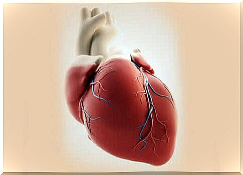
Truncus arteriosus is a birth defect. In addition, a single artery emerges from the heart. In other words, the baby has one large artery instead of two separate ones to carry blood to the lungs and the rest of the body.
This is a fairly rare heart defect. In general, there is an incidence of 0.21-0.34% in the babies born with a congenital heart defect. It is believed that about 2-3% of babies are registered with a pediatric cardiac surgeon.
Causes of a truncus arteriosus
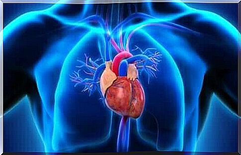
In a normally working heart, the blood follows this cycle: body-heart-lungs-heart-body. When there is a truncus arteriosus, the blood leaving the heart does not flow through the body through the normal route.
In some cases, the heart does not have four separate compartments. Instead, there is only one cavity. So there are no atria or ventricles that separate the blood (that is, depending on its origin and destination).
There is only one common artery. Also, there is no specific route for blood rich in carbon dioxide, nor is there a separate route for the oxygen-rich blood.
A common artery appears during fetal growth as the baby’s heart develops. This should later in development split into 2 separate blood vessels. This condition is thus already present at the time of birth. That is why we speak of a congenital heart defect.
In most cases, the cause of this deviation is still unclear. However, we know of several factors that can increase the risk of a truncus arteriosus. Some of these factors are:
- a family history of congenital heart problems.
- fetuses with chromosome abnormalities are at higher risk of developing this condition. This is especially true for fetuses with velocardiofacial syndrome or DiGeorge’s syndrome.
- certain medications taken during pregnancy can harm the development of a fetus.
- pregnant women who get viral illnesses such as rubella are more likely to have a baby with an abnormality such as truncus arteriosus.
What are the symptoms?
Different symptoms may be seen in each fetus and baby. However, there are some common symptoms that occur in most babies who suffer from this condition. The most common signs are:
- cyanosis
- fatigue
- to sweat
- cold skin
- difficult, accelerated breathing
- increased heartrate
- airway congestion
- lack of appetite
However, all of these symptoms can also occur with other medical conditions or other heart problems. It is therefore very important that the doctor conducts further investigations if a child shows any of these symptoms.
How is it diagnosed?
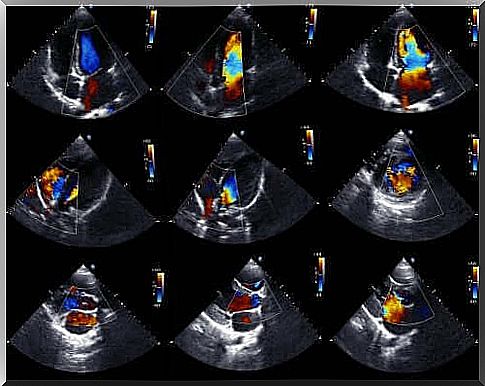
Doctors can usually diagnose these types of anomalies even before a baby is born. To do this, they use a fetal echocardiogram. This is a technique that uses sound waves to reproduce a moving heart image and is one of the most effective techniques for diagnosing congenital heart problems.
Thanks to this research, we can see the appearance of the heart and examine its functioning even while a fetus is still in the womb. With this information, doctors can take steps to administer treatment immediately after birth.
There is also a pulse oximetry test. This is a simple test that measures the amount of oxygen in the bloodstream. A low amount of oxygen in the blood is often the first indication that there is a problem with the functioning of the heart.
Treatment for a truncus arteriosus
Unfortunately, current data on this abnormality indicate that an average of 50% of babies with a truncus arteriosus worldwide die within the first month of life. After this, the survival of the first year of life is between 10-25%.
The vast majority of patients survive the first year of life. Nevertheless, many of them also have serious lung conditions, which are often irreversible.
As far as treatment goes, babies with this condition usually require open heart surgery to both correct the situation and avoid possible complications. This procedure is generally performed during the first month after birth.
During the surgery, the doctor separates the aorta and the pulmonary arteries. This is to create a pathway for blood to flow from the right ventricle to the lungs. They also correct any other detected heart defects where possible.
This operation is not easy, but usually successful. If surgery doesn’t fix this problem, the baby could die.